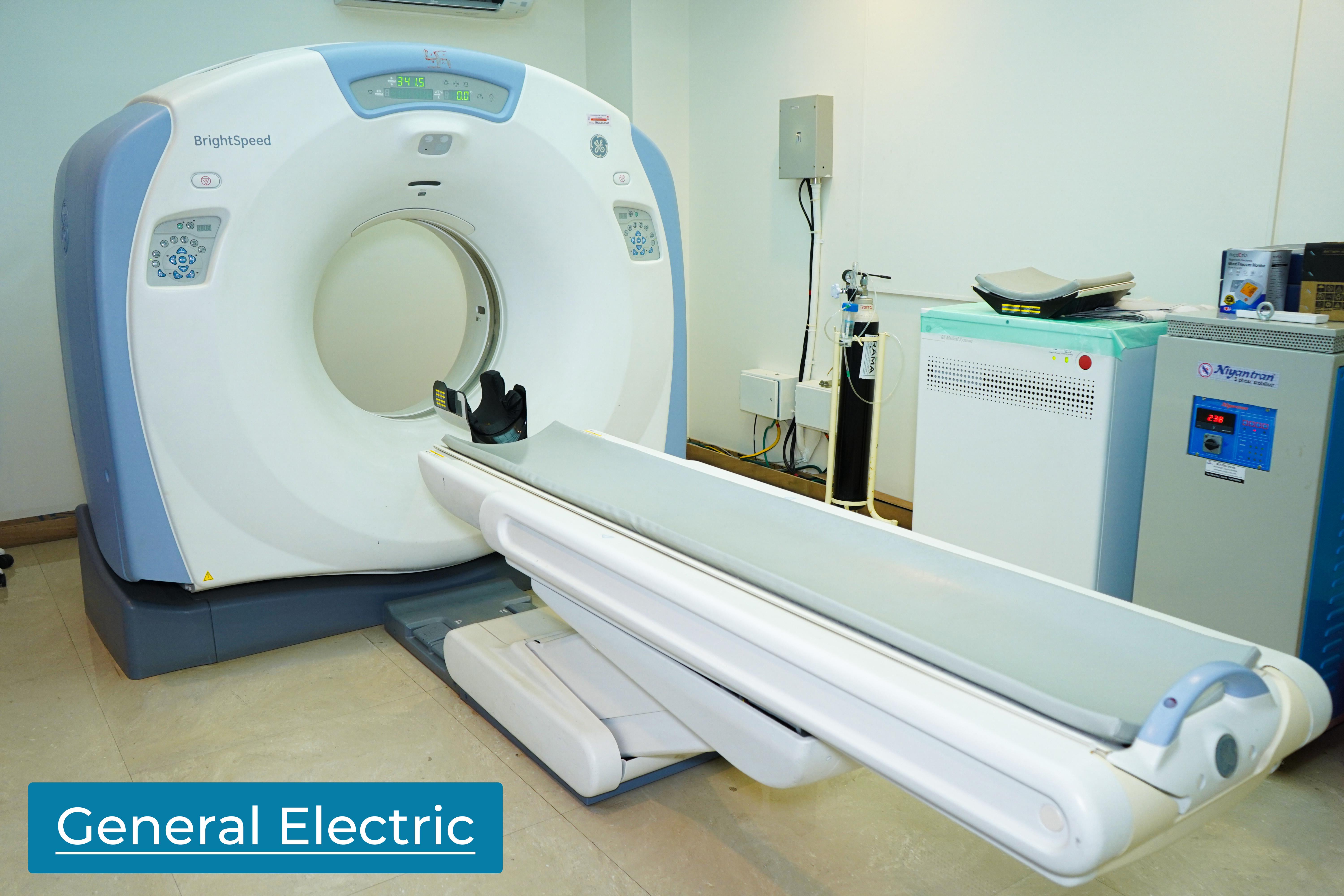
HRCT Chest
HRCT (High-Resolution Computed Tomography) of the chest is a specialized medical imaging technique that provides detailed and high-quality images of the structures within the chest, including the lungs, airways, blood vessels, and surrounding tissues. This diagnostic tool is particularly useful for evaluating various respiratory and pulmonary conditions.

Key Features of HRCT Chest
- High Resolution:HRCT utilizes advanced technology to capture detailed and high-resolution images, allowing for better visualization of small structures within the chest.
- Detailed Lung Assessment:It provides a detailed assessment of lung parenchyma, helping in the diagnosis and monitoring of lung diseases such as interstitial lung disease, pulmonary fibrosis, and infections.
- Evaluation of Airways:HRCT is effective in visualizing the airways, making it useful for diagnosing conditions like bronchiectasis, chronic bronchitis, and bronchiolitis.
- Vascular Imaging:Blood vessels within the chest, including the pulmonary arteries and veins, can be accurately imaged to assess for conditions like pulmonary embolism or vascular abnormalities.
- Detecting Nodules and Masses:HRCT is sensitive in detecting small nodules, masses, or abnormalities in the lung tissue, aiding in the early diagnosis of lung cancer.
- Assessment of Mediastinum:It helps in evaluating the structures in the mediastinum, such as lymph nodes and the heart, assisting in the diagnosis of various mediastinal conditions.
- Quantification of Lung Density:HRCT allows for the quantification of lung density, which is valuable in assessing conditions like emphysema or lung air trapping.
- Follow-Up and Monitoring:HRCT is often used for follow-up and monitoring of treatment response in individuals with known pulmonary conditions.
