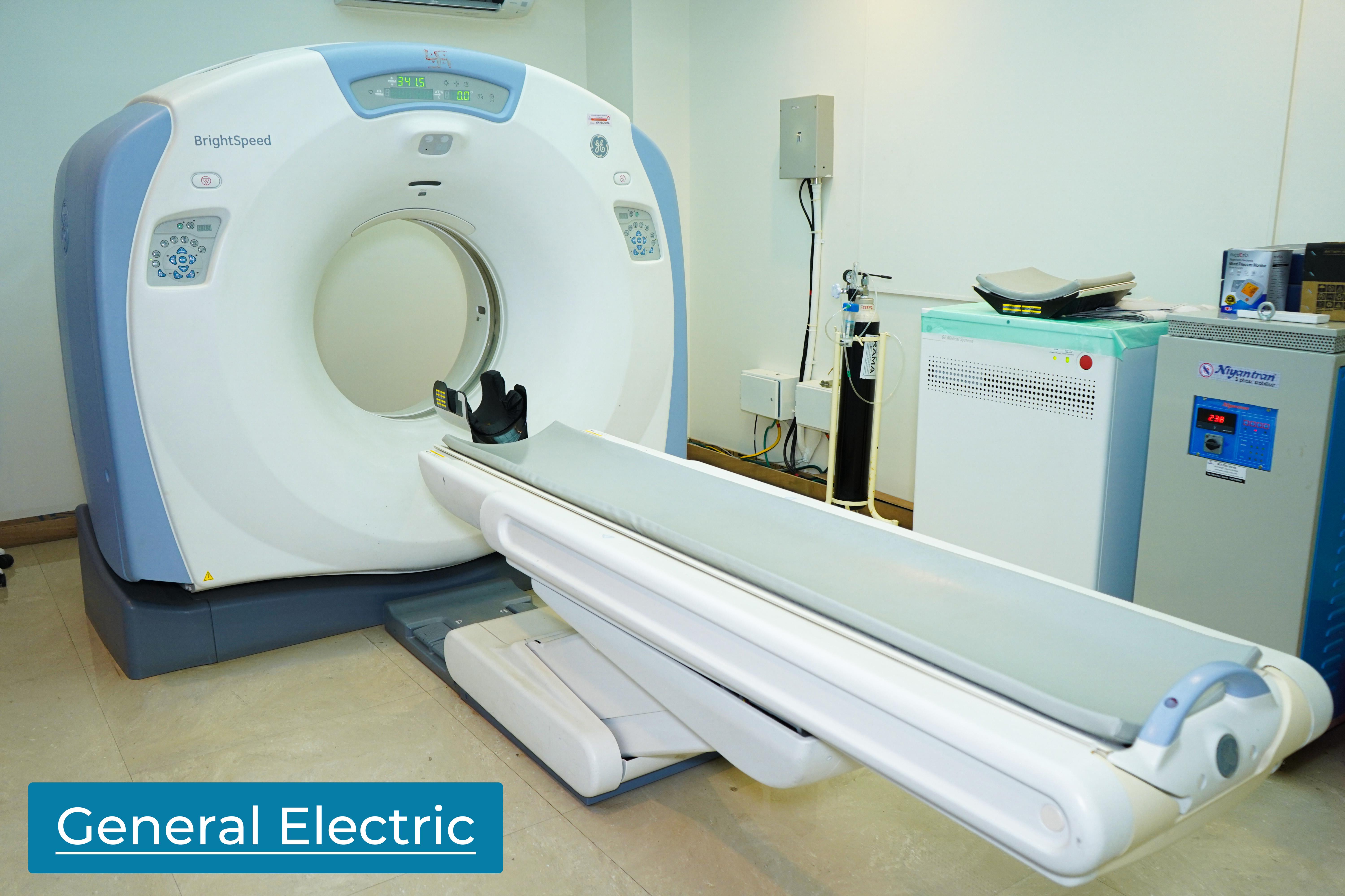
Fistulography:
Fistulography is a diagnostic imaging procedure designed to visualize and assess fistulas, which are abnormal connections or passages between two organs or vessels. This procedure utilizes contrast material and imaging techniques to provide detailed images of the fistulous tract, aiding in diagnosis and treatment planning.

Key Features of Fistulography:
- Identification of Fistulous Tracts:Fistulography is mainly used to recognize and describe the structure of fistulous passages, enabling medical practitioners to determine the scope and position of the unusual connections.
Evaluation of Fistula Origin and Termination:The procedure helps in determining the origin and termination points of the fistula, providing crucial information for surgical or interventional procedures.
Assessment of Underlying Causes:Fistulography helps to determine the root causes of fistulas, such as infections, inflammatory issues, or complications from previous surgeries.
Guidance for Treatment Planning:Fistulography uses detailed images to help healthcare providers plan suitable treatment strategies, whether it be through medical management or surgical intervention.
Real-Time Imaging:During the procedure, live imaging enables the observation of the contrast material moving through the fistulous tract, helping to evaluate its features.
Non-Invasive and Minimally Disruptive:Fistulography is a minimally invasive procedure, usually performed with the introduction of contrast material through a catheter, making it a well-tolerated and relatively low-risk diagnostic method.
